You don’t have to be a cartoonist to have animation be a big part of your life. Animation is everywhere—in our homes, schools, work, and everywhere there’s a screen. And if you grew up in the United States, chances are you’ve witnessed seminal accomplishments in animation history without even knowing it. Does Snow White and the Seven Dwarfs ring a bell? What about Toy Story…or The Flintstones? These works marked important milestones in animation, an art form that continuously challenges its creators to push technology so artists can to bring to life what the mind can imagine. Let’s celebrate it.
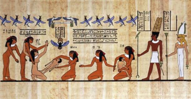
ne of the earliest recorded descriptions of the heart appears in the Ebers Papyrus, an ancient Egyptian medical text written on a scroll nearly sixty feet long. The original ideas are believed to have been recorded around 2600 BC, often attributed to the scholar Imhotep, although the oldest surviving copy dates to about 1700 BC. Within this document, physicians described their understanding of how the heart relates to the rest of the body.
In a section often translated as “The Beginning of the Physician’s Secret: Knowledge of the Heart’s Movement,” the text explains that vessels connect the heart to every part of the body. Ancient physicians believed that by placing their hands on areas such as the head, arms, stomach, or feet, they could examine the heart through these vessels. In this early view, the heart was understood not just as an organ, but as the center of a network of channels that carried life throughout the body.
The Greek physician Galen of Pergamon (129–216 AD) was one of the most influential medical thinkers of the ancient world. Working in Rome, he studied anatomy through animal dissections and carefully recorded how different organs functioned. Nearly two thousand years before modern evolutionary theory, Galen already recognized the close biological similarities between humans and animals, using this idea to guide his medical investigations
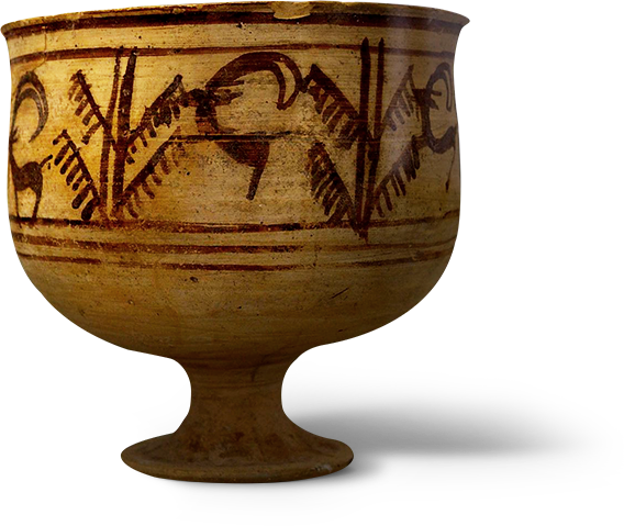
A bronze-age pottery bowl depicts goats leaping
(Shahr-e Sukhteh, Iran).
During the Renaissance, Leonardo da Vinci brought a new way of studying the human body through careful observation and detailed drawings. Fascinated by anatomy, he dissected human and animal hearts to understand their structure and function. His sketches revealed the chambers of the heart, the twisting pattern of the heart muscle, and the way the valves open and close as blood flows through them.
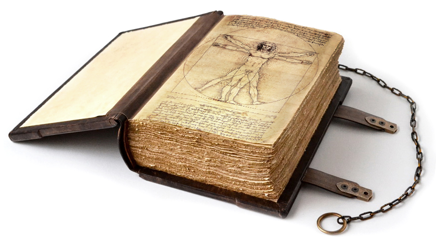
Through experiments with water and glass models, Leonardo even studied how fluid moves through the heart’s valves, anticipating ideas about blood flow that would not be fully understood for centuries. Although his anatomical notebooks remained unpublished during his lifetime, his work represents one of the earliest attempts to study the heart through direct observation and scientific illustration.
Between the Renaissance and the rise of modern cardiology, progress in understanding the heart moved slowly but steadily. Physicians began to observe the heart more carefully through anatomy, experimentation, and new medical tools.
English physician William Harvey published De Motu Cordis in 1628, demonstrating that the heart functions as a pump that circulates blood throughout the body. Through careful observation and experiments, he showed that blood moves in a continuous loop rather than being consumed by the body as previously believed. Harvey’s discovery established the modern concept of circulation, becoming one of the most important foundations of cardiovascular science.

French physician René Laennec invented the stethoscope in 1816, allowing doctors to listen to heart and lung sounds without placing their ear directly on the patient’s chest. This simple instrument revolutionized clinical medicine, enabling physicians to detect abnormal heart sounds, valve problems, and irregular rhythms. The stethoscope became one of the most important tools for diagnosing heart disease.

A bronze-age pottery bowl depicts goats leaping
(Shahr-e Sukhteh, Iran).

Musical Mews and Feline Follies introduced Felix the Cat—often
considered the first animated movie star.
In the 1940s, pediatric cardiologist Helen B. Taussig helped transform the treatment of congenital heart disease. While working at Johns Hopkins, she cared for infants with Tetralogy of Fallot, a condition that left them oxygen-deprived and known as “blue babies.” Taussig proposed increasing blood flow to the lungs, which led to the creation of the Blalock–Taussig shunt in 1944. This groundbreaking procedure became one of the first successful surgeries for congenital heart defects and marked the beginning of modern pediatric cardiology.
In 1947–1948, American surgeon Claude Beck performed the first successful cardiac defibrillation during surgery. A young patient’s heart had stopped due to ventricular fibrillation, and Beck used an electrical shock applied directly to the heart to restore a normal rhythm.This moment proved that the heart could be restarted electrically, opening the path for modern defibrillators and resuscitation techniques.
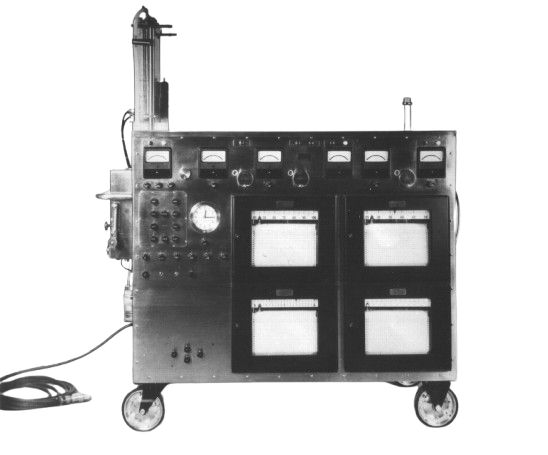
In the early twentieth century, American surgeon John H. Gibbon Jr. developed a device that would transform heart surgery: the heart–lung machine. While assisting in surgery for a patient with a pulmonary embolism in 1931, Gibbon wondered if a machine could temporarily take over the work of the heart and lungs, allowing surgeons to operate on the heart itself.After more than two decades of research and experimentation, Gibbon successfully used his machine in 1953 to repair an atrial septal defect. By circulating and oxygenating the patient’s blood outside the body, the heart–lung machine made it possible to stop the heart while maintaining blood flow. This breakthrough opened the door to modern open-heart surgery, allowing physicians to repair conditions that were once considered impossible to treat.

The thaumatrope housed a rotating mechanism with a different
picture on each side.
When rotated, you saw a combined picture (known as persistence
of vision).

The phenakitoscope featured spinning disks reflected in
mirrors that
made it seem like the pictures were moving.
The zoetrope was a hollow drum that housed images on long interchangeable
strips that spin and made the images appear to move.
The flip-book, also known as the kineograph, reached a wide
audience and is credited
with inspiring early animators more than the machines
developed in this era.

The praxinoscope expanded on the zoetrope, using multiple
wheels to rotate images.
It is considered to have shown the first prototypes of
the animated cartoon.
A bold experiment that changed medicine forever. Werner Forssmann became the first person to catheterize a living human heart—his own.
Werner Forssmann was only twenty-five when he began imagining a way to reach the human heart from the inside. While working as a young physician at Auguste-Viktoria Hospital in Germany, he often performed autopsies and noticed that although the body could appear intact, the heart frequently told the real story of death—damaged valves, malformed chambers, and blocked vessels. At the time, heart disease was something doctors could only observe after death, not treat in the living. Inspired by earlier veterinary experiments, Forssmann believed a thin tube could be inserted through a vein and guided safely to the heart, allowing doctors to diagnose and eventually treat cardiac disease. Many physicians considered the idea too dangerous, but Forssmann was determined to prove otherwise.
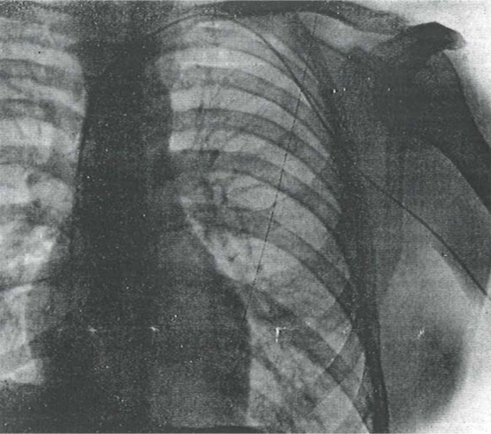
In 1929, after receiving permission to experiment only on animals, Forssmann decided to test the idea on himself. With the help of an operating room nurse, Gerda Ditzen, he secretly inserted a catheter into a vein in his own left arm and carefully advanced it toward his heart. To confirm the experiment, he walked two flights of stairs to the radiology department with the catheter still inside his body and captured an X-ray showing the tube reaching his heart. That image became the first successful demonstration of cardiac catheterization in a living human. Though risky at the time, Forssmann’s daring experiment opened the door to modern cardiology, making it possible for physicians to explore, diagnose, and treat the heart from within.
orssmann’s life after his daring experiment was anything but easy. Although he later worked at the Charité Hospital in Berlin, controversy surrounding his early publication cost him his position, and many physicians dismissed his work as reckless. Soon after, World War II began and Forssmann was sent to the front as a military medic. Meanwhile in the United States, André Cournand and Dickinson Richards rediscovered and expanded his idea of cardiac catheterization, proving its value in studying the heart. By the time the war ended, the technique he had demonstrated was already gaining recognition abroad, while Forssmann struggled to rebuild his life in Germany and eventually worked quietly as a urologist in the town of Bad Kreuznach.
One day after finishing routine surgeries, Forssmann learned surprising news: the Nobel Prize in Physiology or Medicine had been awarded for the development of cardiac catheterization. Moments later, the hospital director confirmed that he himself was one of the recipients, sharing the prize with Cournand and Richards. The experiment that once nearly ruined his career had finally been recognized for transforming cardiology. Forssmann later retired from medicine in 1969, and ten years afterward he died of a heart attack. Today, the hospital where he performed his bold experiment bears his name, honoring the man who first dared to reach the human heart from within.